Hair Root Under Microscope
There are many variations among horses in hair colour - especially the colours we register.

Hair root under microscope. Hair is composed of a strong structural protein called keratin. • Straight hair tends to be round in shape. Download 1,312 Hair Microscope Stock Photos for FREE or amazingly low rates!.
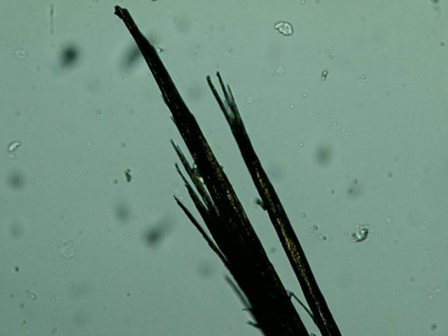
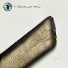
Either physiological or emotional stress can be the root cause of diffuse hair loss. So I took a pair of scissors and cut one strand of hair so it would have a fresh cut end and put it under the microscope, hoping to find some differences to a piece I just pulled from my head. A bat hair (c).
Though it often appears soft and smooth to the eye, especially from a distance, deer hair is relatively coarse. Mammalian hair is composed of a protein, keratin. Hair that has been pulled out from the root can grow back under which of the following circumstances?.
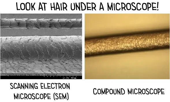
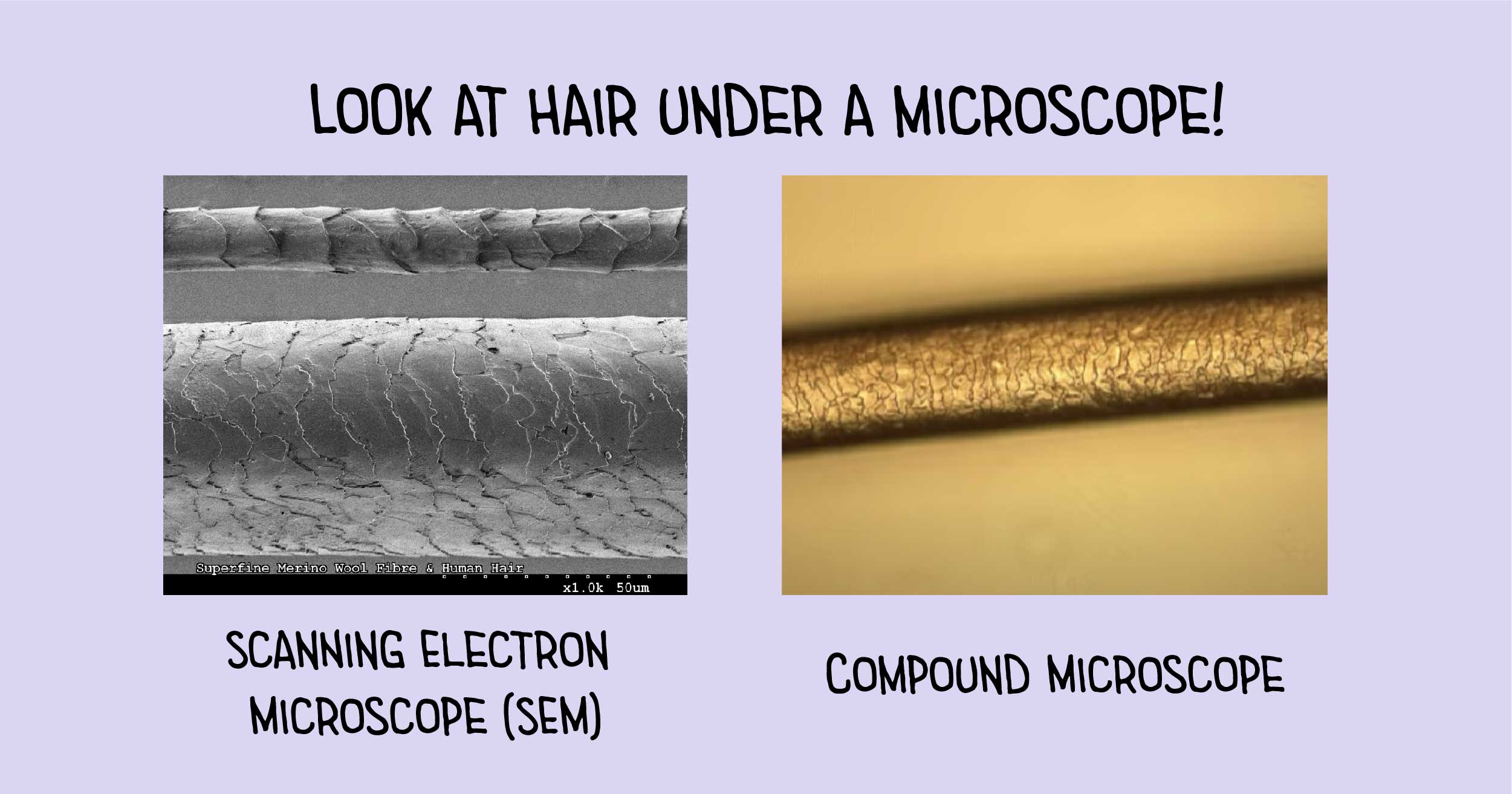
A close-up of a part of male skin with partially shaved facial hair with a strong magnification under a microscope. Hair Under a Compound Microscope Make a thin layer of nail polish at the central part of a clean glass slide Using a brush, brush over the nail polish to create an even thin layer of the nail polish Using the tweezers, place one or several strands of hair on the central part of the slide with nail. Find Root Hairs Cells Under Microscopic View stock images in HD and millions of other royalty-free stock photos, illustrations and vectors in the Shutterstock collection.
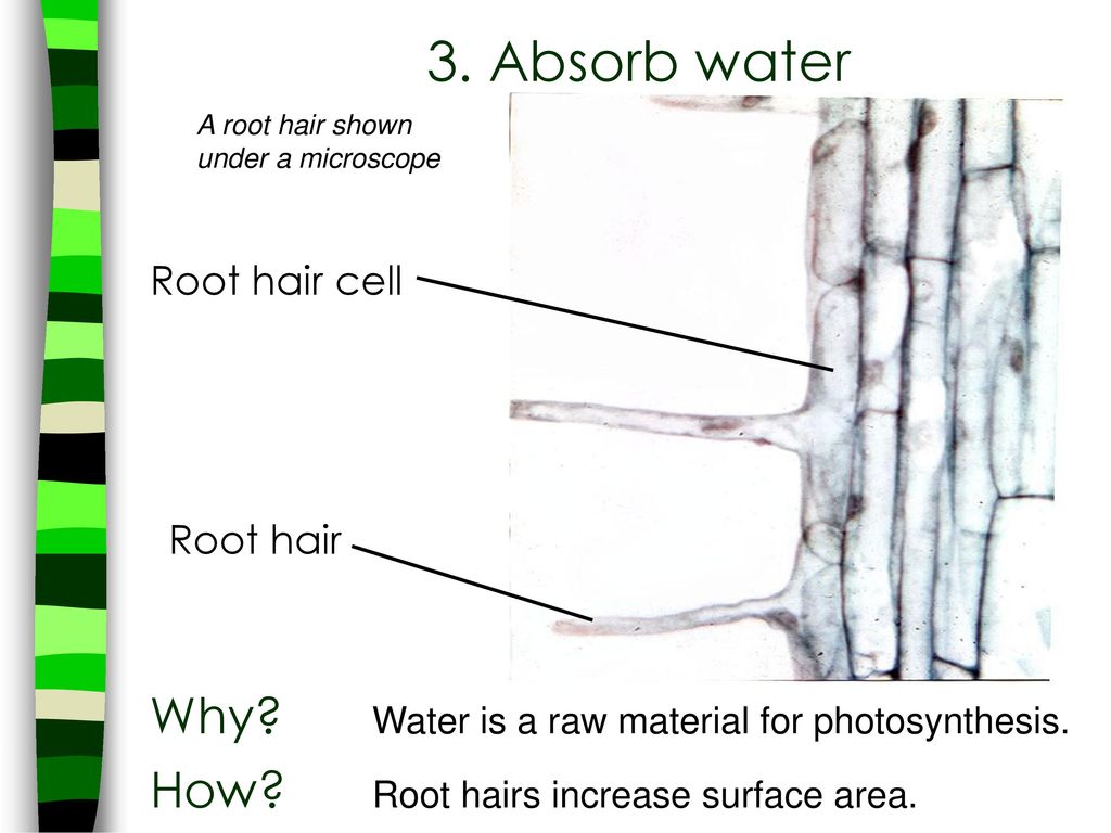
The scales are made of the same, tough keratin protein that is found in skin, fingernails and toenails. Zoomed under the nano microscope. We should mount this root on a microscope slide and add a drop of water.
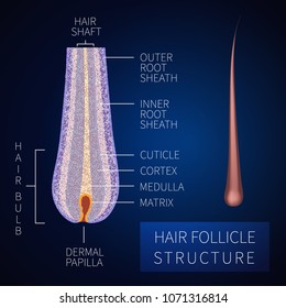
This can help determine whether an infection is causing hair loss. As a result, there is a lack of adhesion between the hair shaft and the root sheath, and the hair fiber is poorly anchored in the follicle. In the hair bulb, living cells divide and grow to build the hair shaft.
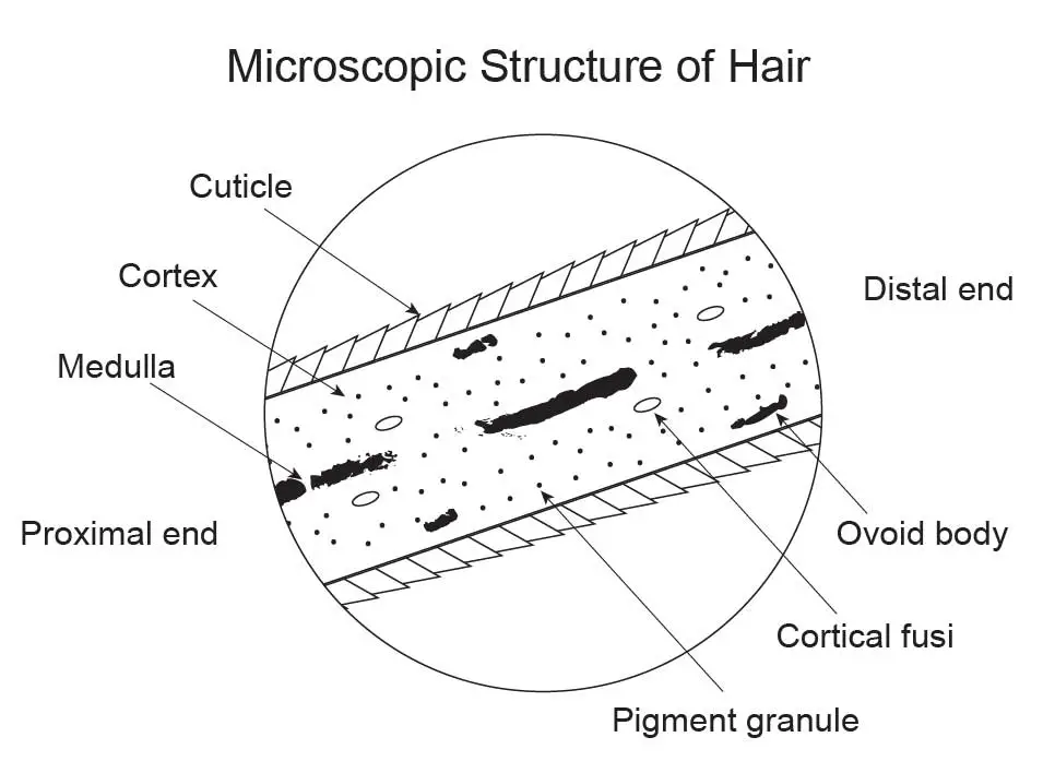
There is a gradual conversion from terminal hair follicles to vellus-like follicles in clients with:. Mammalian hair consists of three distinct morphological units, the cuticle, the cortex and the medulla. By comparison, the success rate of microscopic root canal treatment (performed with the help of an operating microscope) is more than 90 percent.
Human hair under microscope illustration. A study of 99 adult male and female rhesus macaques sought to examine the relationship between hair loss and chronic hypothalamic–pituitary–adrenal (HPA) axis activity by. These flat scales are called the cuticle;.
By hoofpick August 3, 11 Updated April 23, 13. New comments cannot be posted and votes cannot be cast. Image of medicine, root, microscope -.
Bacteria preferentially target hair roots that were actively growing at time of death. Human gray hair on black background. At the end of the root is a thickened bulb that works in conjunction with the underlying papillae to facilitate the nourishment, development, and growth of the hair.
Stem and root under a microscope. Horse Hair Under a Microscope:. Posted by catcaro on January 11, 16.
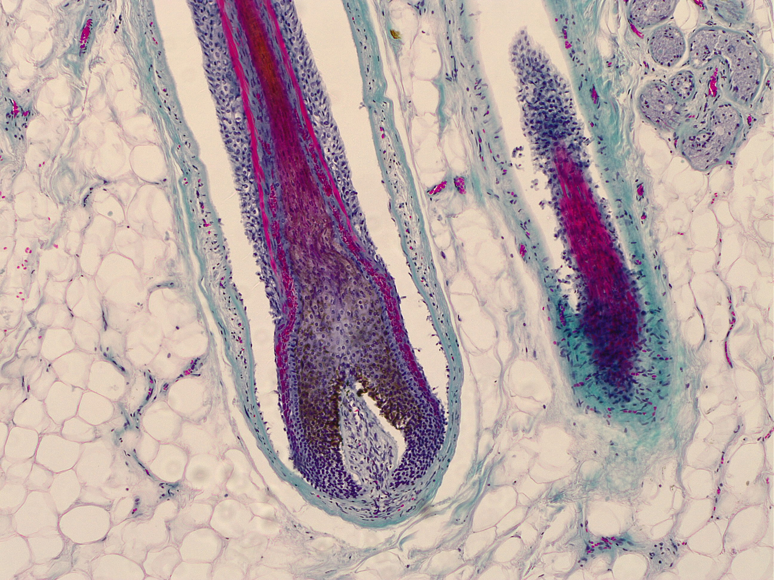
Histology of human tissue, show skin with hair follicles as seen under the microscope. Photo about Human Hair root seen on microscope at 100x magnification. The particular hue (colour shade), value (lightness or darkness), and intensity (saturation) of a specimen are enhanced through microscopy so that.
(v) Name the pressure that helps in the movement of water. Tions in the root diameter of field-grown plants under a microscope without washing. Histology of human tissue, show skin with hair follicles as seen under the microscope.
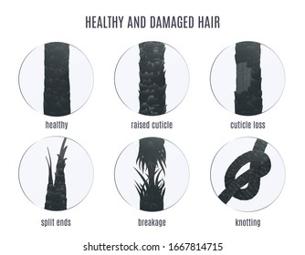
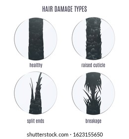
Sential that a comparison microscope be utilized for this stage of the examina. Ture under transmitted light or a white structure under reflected light. This is caused by premature breakage, typically a result of tension or physical stress.
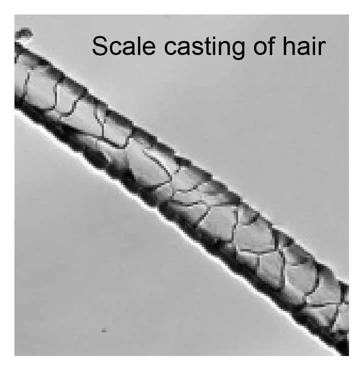
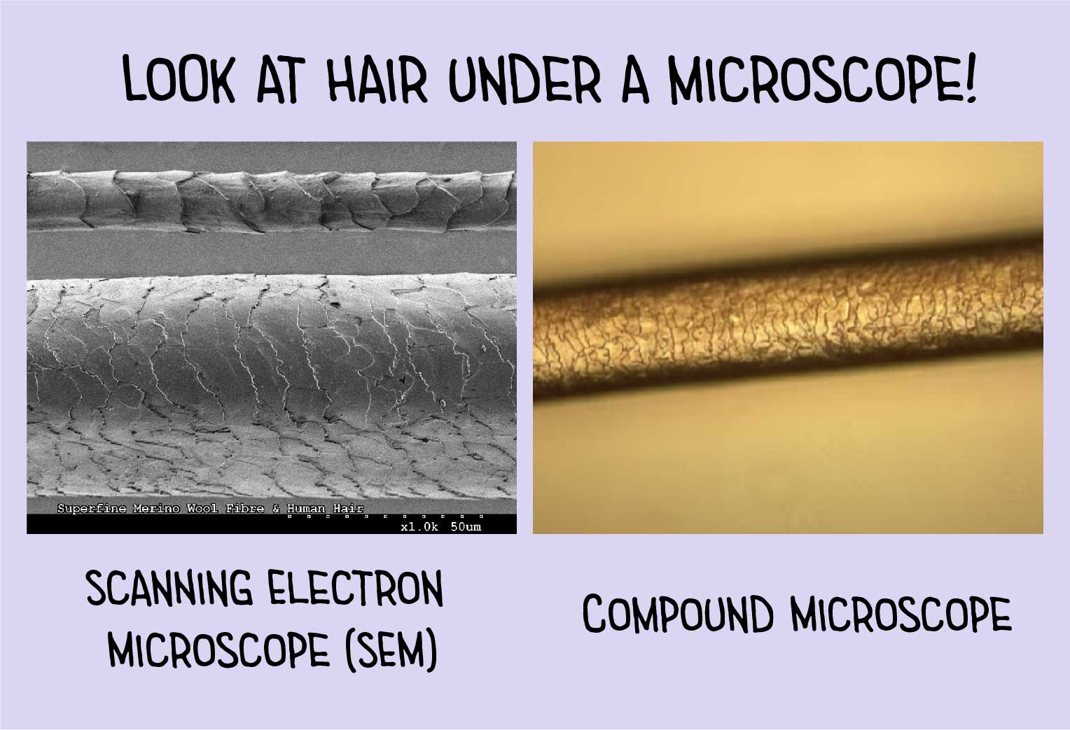
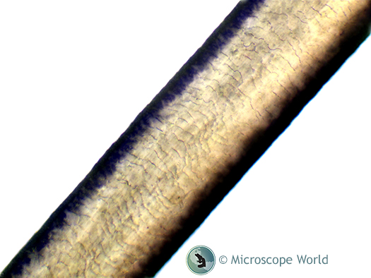
Examination of hair cuticle scale patterns It is very difficult to directly observe scale pattern from hair strand on a slide. (iii) Under what conditions in the soil will the root hair cell resemble :. A single human hair under intense magnification.
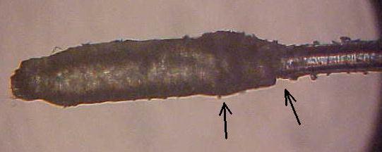
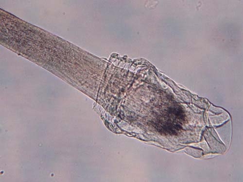
Mycelial threads, large and branching, are often seenwithin the hair. This thread is archived. Each hair on the human scalp is divided into a hair shaft that protrudes from the skin and a hair root, which is embedded in the skin.
Cell B was placed before being viewed under the microscope. Hair microscopy is used to view the end of a hair and establish whether it contains a root. This parasite appears under the microscope chiefly in the form of a largenumber of round spores, irregularly grouped or massed about the follicularportions of the hair.
Examine the hair shaft under a microscope. Under a microscope, scalp hair and pubic hair reveal a greater range of characteristics than other kinds of human hair, so we use them more often in forensic comparisons. The hair bulb forms the base of the hair follicle.
In addition to these units pigment bodies and other inclusions can help distinguish one hair type from. This helps determine the stage of the shedding process. Conventional root canal treatment is successful in 50 to 60 percent of cases.
For my education as well as yours!. When observed under the microscope most of a hair. The kinks closer to the roots were too ‘young’ so they seemed healthy.
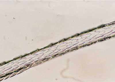
The cortex can also be seen under a microscope to compare the appearance of the pigment granules. The materials you may need:. Microscopic inspections have shown that the hair can be round, oval, flat, and even triangular, as well as many variations of these shapes.
The hair shaft is composed of three layers:. Leave a comment if you have any wisdom to share!. The software that is included with the microscope camera we used allows single snapshots, like the three shown above, and also extended depth of focus images, which essentially combines many single in-focus pictures into one.
Thousands of new, high-quality pictures added every day. Under a high power compound light microscope, the root seems to be made up of numerous cells that are regularly arranged and in different shapes and sizes. Hair-degrading fungus comes under the microscope.
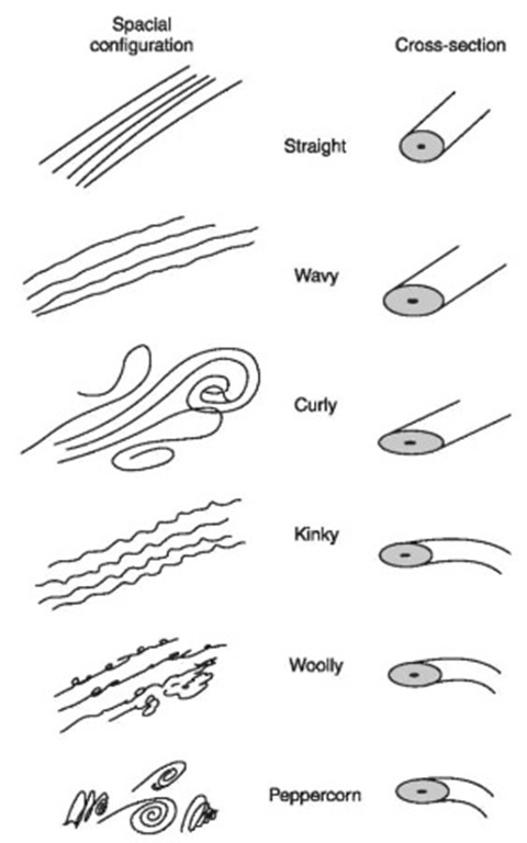
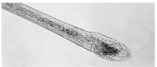
The fur of many young deer often feature white spotting as well. The root of human hairs is commonly club-shaped, whereas the roots of animal hairs are highly variable between animals. Of course, different wave patterns do have more common shapes associated with them, and these are as follows:.
Make sure it's cons. A Visual Exploration of Medicinal Sativa and C. If the hair shaft is from a suspect, these granules can tell the investigator the color of the hair.
So for those of you who think that natural hair does not require protein treatments, think again!. The proximal or root end of the hair to the distal or tip end of the hair (Fig. Looked at my hair under a microscope and my gray hair is actually clear.
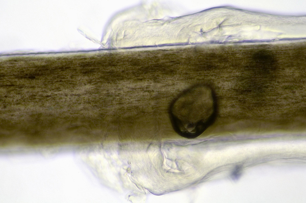
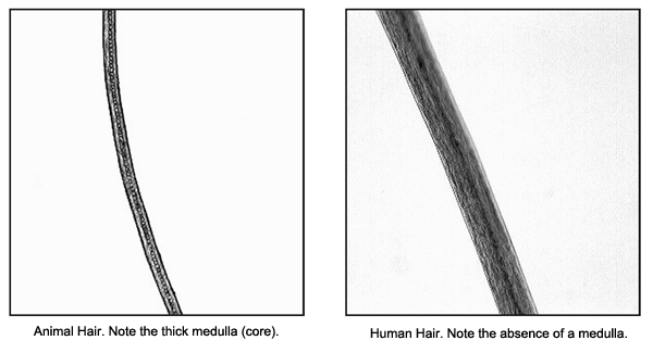
The medulla in animal hairs is normally continuous and structured and generally occupies an area of greater than one-third the overall diameter of the hair shaft. Under a microscope, the appearance of various components of the hair shaft offers lots of information as to who it came from. This is Watson, and today we are going to look at some cool objects under the microscope!.
The bacteria invade the hair shaft near the root. It is the same protein that makes horn, fingernails, claws, skin epithelium, and dander. Deer pelts are usually tawny or brown in color and are often highlighted with white patches along the throat and chest.
The problem of underestimation of root quantification by washing roots during sample preparation will be dis cussed. Hair root follicle or bulb by microscope. Cuticle or outer layer has overlapping scales with free ends.
If so, the. The cortex has not been destroyed. A hair follicle anchors each hair into the skin.
Roots and root hairs were exposed through careful separation from the soil mass and all the variation in roots were directly detected. Hair transplant surgeons place a high importance on the integrity of naturally occurring hair follicle units to find improved methods of hair graft harvesting, dissection, grading and transplantation. (iv) Name the pressure responsible for the movement of water from the root hair cell to the xylem of the root.
Hair Loss Under the Microscope. Indica/Ford McCann So yeah, you could say there's more to marijuana. However, the bulb is always present when it comes from the source (that is, the follicle).
134,1,579 stock photos online. Under low-power microscopic examination, a human scalp hair is seen to have an outer layer of flat scales pointing outward from root to tip. It may be small or large, white or pigmented.
Photo about Microscopic view of plant cells for botanic education. Image of microscopy, definition, high -. Your doctor scrapes samples from the skin or from a few hairs plucked from the scalp to examine the hair roots under a microscope.
Originally posted 19:00:21. Photomicrograph of Human Hair Root. The hair was scanned along the length at 10X, 25X, and 40X under an optical microscope to observe the morphological characteristic of the tip and root.
• Wavy hair tends to be oval. Ask questions about family history. For more information on microscope.
January 25, 18 - 06:55. This video is a follow-up to a previous video on hair:. The hair root is lodged in the hair follicle.
It can’t identify specific individuals, but can identify the hair species. To start with our microscope experiment, we first need to get a seedling of a grass with roots that contain a few hairs. Hair shaft under the hair analysis microscope at 400x, focused from the top of the shaft.
If the hair root has tissue, DNA testing can provide absolute identity. Click here to read more articles in the Hair Under The Microscope Series. It was accepted as a forensic science by the 1950s.
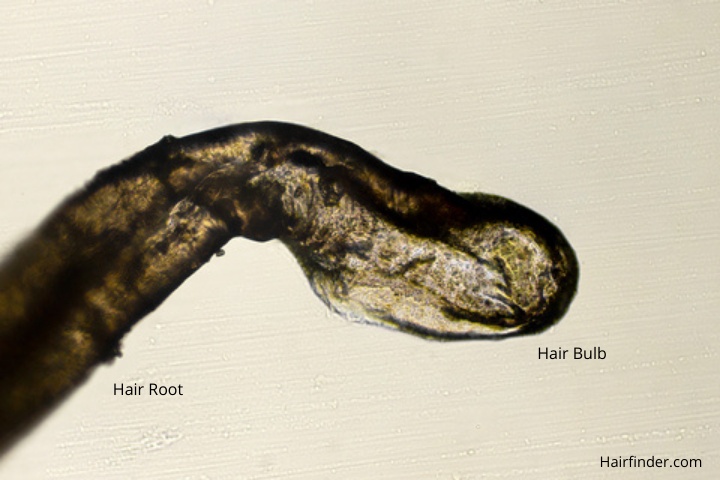
The root of hair ends in an enlargement (referred to as a hair bulb) that is whiter in color and softer in texture than the shaft of hair. How is it set up ?. A postmortem root band (PMRB) is an opaque microscopic band that can be observed near the root area of hairs from a decomposing body.
Check out Part 2 (We look. Looks like a large metal cable a giant tree root cluster, who would have guessed. New users enjoy 60% OFF.
Microscopic hair analysis is the science of comparing several strands of hair under a microscope and attempting to deduce if the strands 'match'. All hair lost at the root will contain a bulb. Forensic scientists perform 3 major types of hair analysis:.
I've had beard hair where half the shaft is white, and the part closer to the root is black. Your doctor gently pulls several dozen hairs to see how many come out. Kateryna Kon / Shutterstock.
Researchers often monitored more than a dozen attributes, including pigment distribution and scale patterns. Composed of four stacked exposures to achieve greater field. Hair root captured using the Jenoptik C14+ microscope camera.
Microscope slides Mounting media (it could be water or 70% glycerol wet mount medium) Coverslips Tweezers or forceps Different hair specimens A compound microscope. Image courtesy th US edition of Gray's Anatomy of the Human Body. Scanning electron microscope image of cannabis budCannabis Under the Microscope:.
A descriptive report.
Fbi Deedrick Forensic Science Communications January 04

Texas Cryptid Hunter Alleged Black Panther Hair Analysis
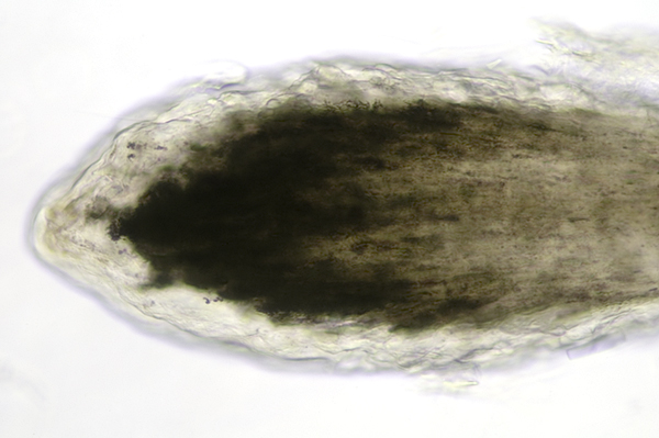
Mic Uk Hair Through The Microscope
Hair Root Under Microscope のギャラリー

Microscopic Hair Comparison Sciencedirect

Alfalfa Root Tip Revealed By Scanning Electron Microscopy A View Of Download Scientific Diagram
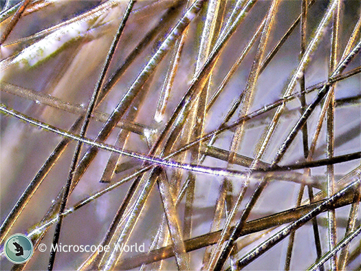
Hair Analysis Under The Microscope
Fbi Deedrick Forensic Science Communications January 04
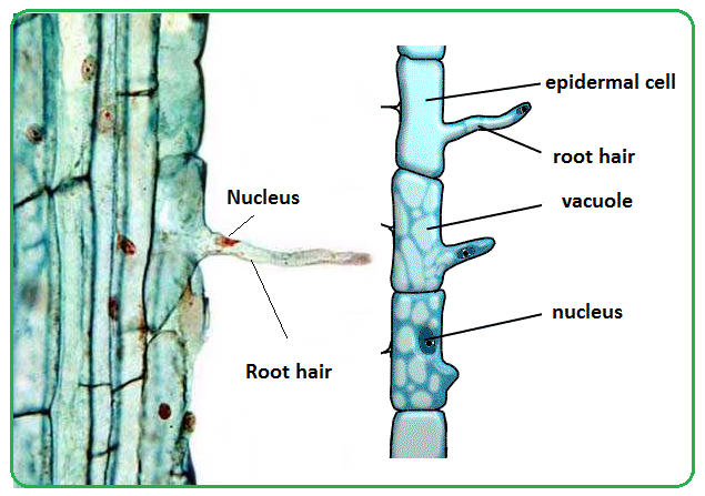
Biology Notes For Igcse 14 61 Root Hairs And Water Uptake By Plants
Untangling A Hairy Science

111 19 Megan Lim
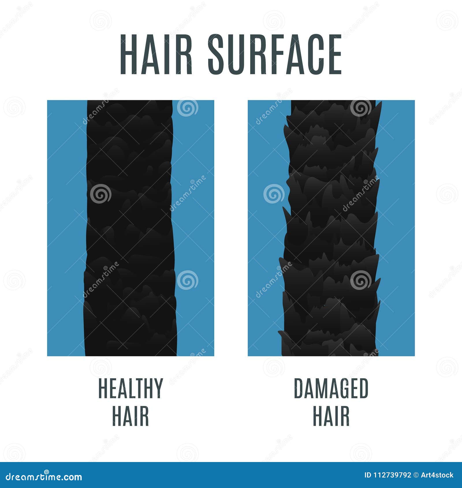
Healthy And Damaged Hair Surface Stock Vector Illustration Of Shampoo Root
Mic Uk Hair Through The Microscope
Dog Hair Under The Microscope Steemit
Under The Microscope Get Forensic With Hair Analysis Carolina Com

Entire Hairs Plucked Under A Microscope Soooooo Awesome Youtube

Horse Hair Under A Microscope A Descriptive Report Color Genetics

Pin On Hair N Metal

Root Hair Cells Black Arrow Pointing At One Of The Root Hair Cells Download Scientific Diagram
Embedded Hair Roots Under 10 Magnification Optical Digital Microscopy Download Scientific Diagram

Tissue Examination Histologyolm
Root Hair Microscope High Resolution Stock Photography And Images Alamy
What Is Hair Made Of And How Does It Grow
Untangling A Hairy Science
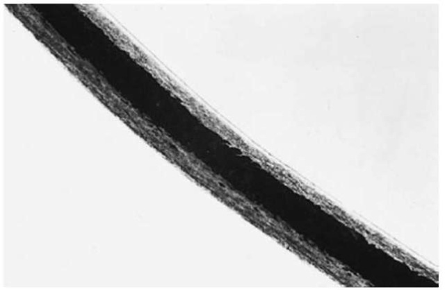
Identification Of Human And Animal Hair
Comparison Microscopic

The Arabidopsis Rab Gtpase Raba4b Localizes To The Tips Of Growing Root Hair Cells Plant Cell
Damaged Human Hair Under The Microscope Steemit

Rat Guard Hair Near Root Under The Microscope

Identification Of Human And Animal Hair
Hair Under A Microscope Rs Science
Hair Under A Microscope Rs Science

Hair Being Examined Under Microscope Interactive Investigator
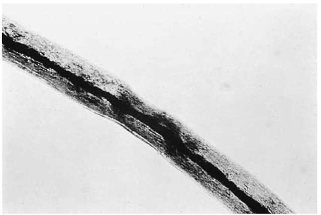
Identification Of Human And Animal Hair

Root Hairs Youtube
3

Hair Under The Microscope Compound And Stereo Microscope Observations
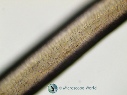
Hair Analysis Under The Microscope

Hair Follicle Structure Microscopic Anatomy Of A Hair Follicle In Its Download Scientific Diagram
Damaged Human Hair Under The Microscope Steemit
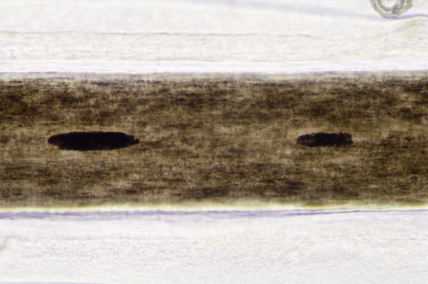
Mic Uk Hair Through The Microscope
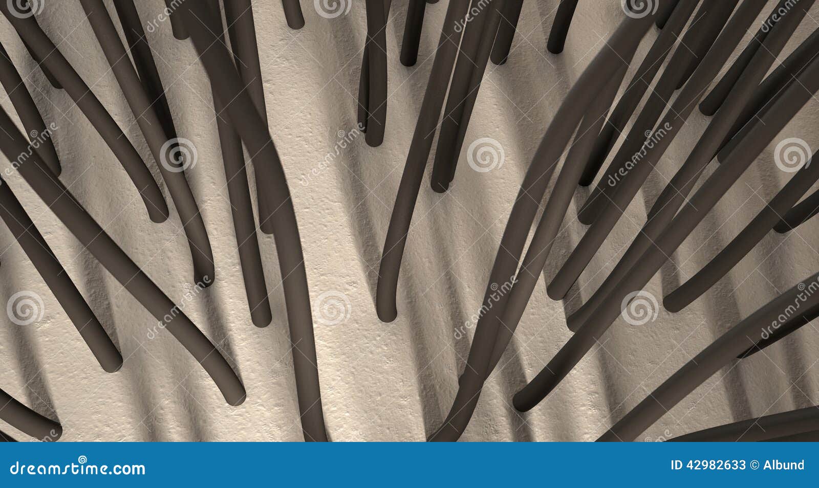
Microscopic Hair Roots Stock Image Image Of Fiber Research

Science Source Dog Hair With Root

File Human Hair Root03 Jpg Wikimedia Commons
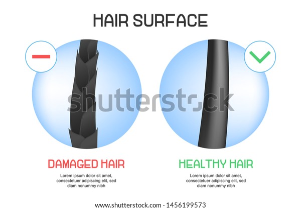
Surface Healthy Damaged Hair Under Microscope Stock Vector Royalty Free
3
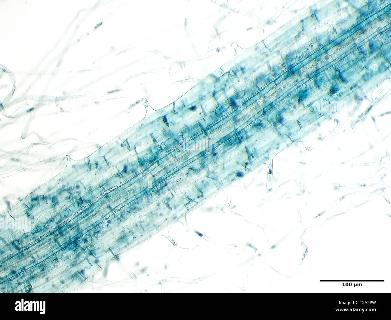
Microscopic Hair High Resolution Stock Photography And Images Alamy

B Schematic Of A Terminal Human Hair Follicle And Its Sebaceous Download Scientific Diagram

A Hair Mystery Curly Hair Gone Straight Cabelos Saudaveis Cabelo Cabelo Encaracolado

Horse Hair Under A Microscope A Descriptive Report Color Genetics
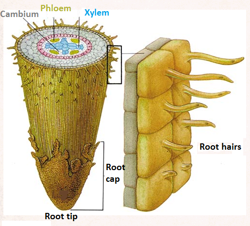
Root Hairs And Water Uptake By Plants Biology Notes For Igcse 14
Untangling A Hairy Science
The Microscope As A Tool In Tanning And Taxidermy

Kori Beckham Hair Analysis Lessons Tes Teach
Hair Follicle Microscope Images Stock Photos Vectors Shutterstock

Hair Follicles Grafts Under Microscope Youtube
Q Tbn 3aand9gcr4e Aopysou3n7zcftagbcx2eeepcxl3w Ddzz7kwiskssf5cj Usqp Cau

Hair Strand Under Microscope

Squishy S 2 Ingrown Hair Root Removal Microscopic Youtube
Hair Under A Microscope Rs Science

Cress Root Tip With Root Hairs Microscope Pictures Microscopic Pictures
Forensic Science Hair
Hair Follicle Microscope Images Stock Photos Vectors Shutterstock

Under The Microscope Hair Follicle Nerve Endings
Hair Under A Microscope Rs Science
Untangling A Hairy Science

Photomicrograph Of Human Hair Root The Scale Pattern Of The Cuticle In Download Scientific Diagram

Root Hairs Near Tip Of Tomato Root Institute Of Biotechnology
Hair Analysis Under The Microscope
Damaged Human Hair Under The Microscope Steemit
Your Hair Under The Microscope Southland Soap
Hair Analysis Under The Microscope

Hair Biology For Majors Ii
Http Cosmosforschools Com Pdfs Lesson 070 Handout Pdf

Close Up Of Hair Grafts Dissection Under Microscope Youtube
Identification Of Human And Animal Hair
Hair Under A Microscope Rs Science

Cell Proliferation And Hair Tip Growth In The Arabidopsis Root Are Under Mechanistically Different Forms Of Redox Control Pnas
Hair Follicle Microscope Images Stock Photos Vectors Shutterstock
Fbi Deedrick Forensic Science Communications January 04
A Leaf In Time Library Activity Ppt Download

Microscope World Stereo Microscope Microscopic World
Microscope Styled Close Up View Of Human Skin And Hair Roots Stock Photo Alamy
Q Tbn 3aand9gcsbzlr7mu1yz0ksckpexnn033ctlmwbtpwvx0 Geusmga8mmk9v Usqp Cau

Pin On Microscopic

Photomicrograph Of Guard Hair From Lumbar Region Of Different Domestic Download Scientific Diagram
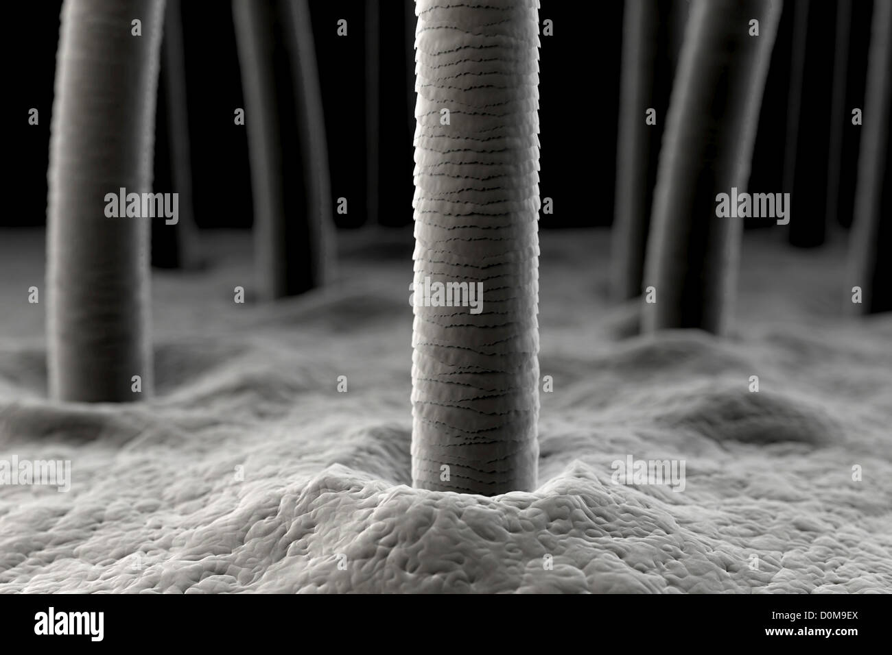
Microscope Styled Close Up View Of Human Skin And Hair Roots Stock Photo Alamy
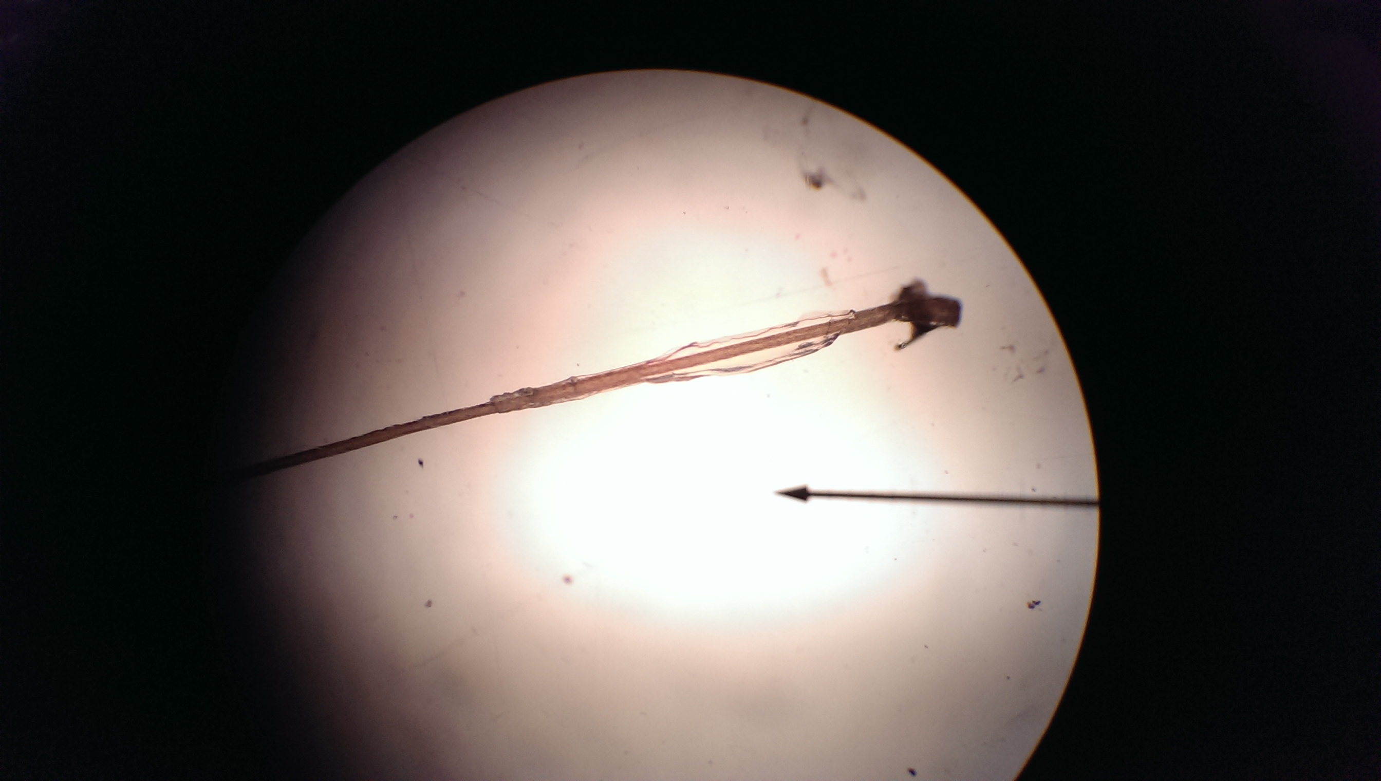
Microscope Lab Report Biolabreports

Hair Analysis Csimodule

Hair Under The Microscope

Vicia Faba Young Root Hair Region Cell Under A Microscope Things Under A Microscope Exam Papers Root

Gallery For Hair Root Microscope Roots Hair Hair Shows Hair Removal Permanent
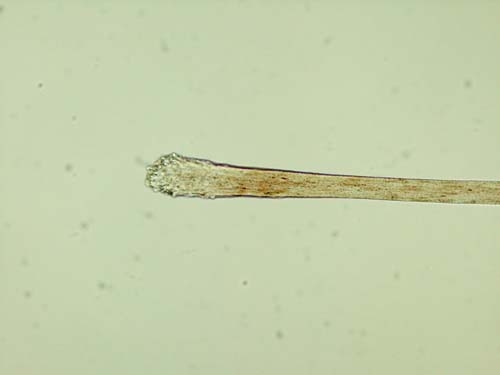
Forensic Science Hair
Buy Human Hair Root Microscope Limit Discounts 51 Off
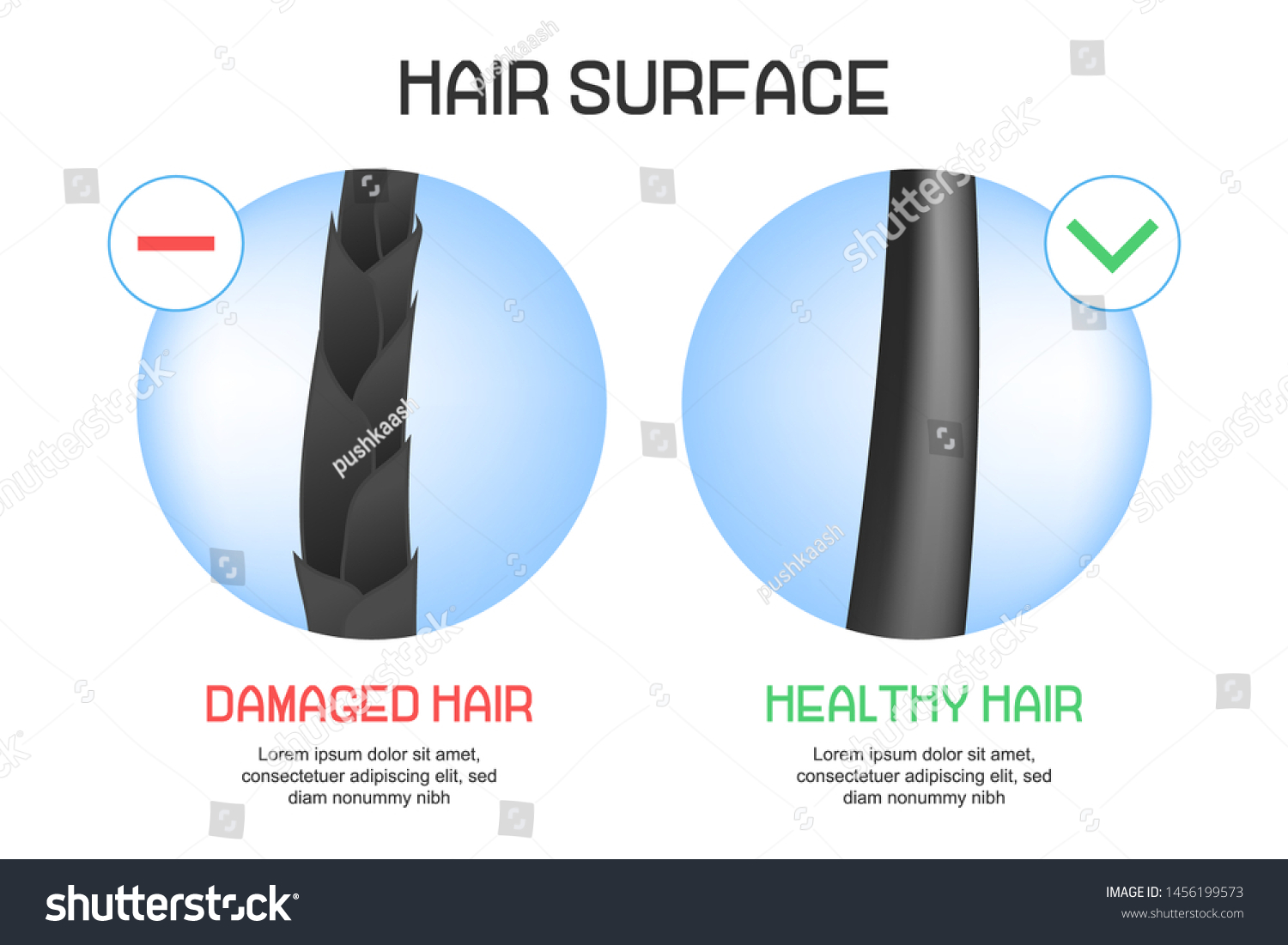
Surface Healthy Damaged Hair Under Microscope Stock Vector Royalty Free

Squishy Slimy Ingrown Hair And Roots Plucked Under A Microscope Youtube

Untangling A Hairy Science

Microscopy Images Of Root Hairs Of Lentil Lupine Wheat And Maize Download Scientific Diagram

Photomicrograph Of Cat Hair Near The Root Cat Hair Cats Root
25 Lovely Dog Hair Under Microscope Demodectic Mange
Some Melanomas May Start In Hair Follicles National Institutes Of Health Nih

Scanning And Light Microscopy Of Alfalfa Root Hair Initiation A Download Scientific Diagram

Trichorrhexis Nodosa Wikipedia

Microscope World Blog Hair Root

Root Hairs W M Microscope Slide Biology Scichem


